|
|
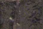
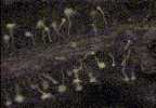
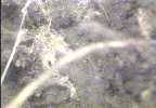
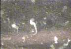
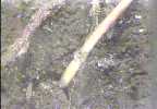
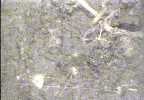
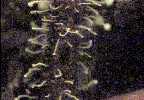
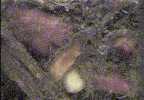
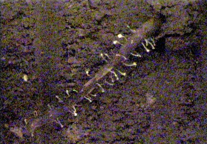
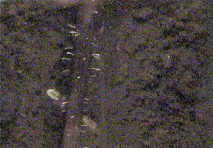
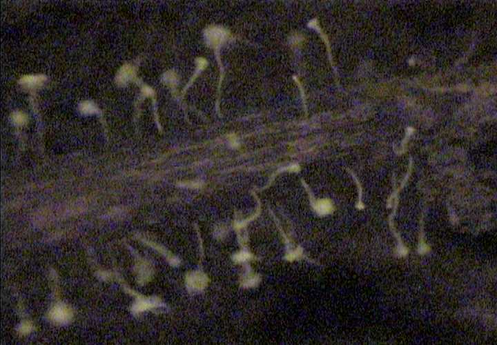
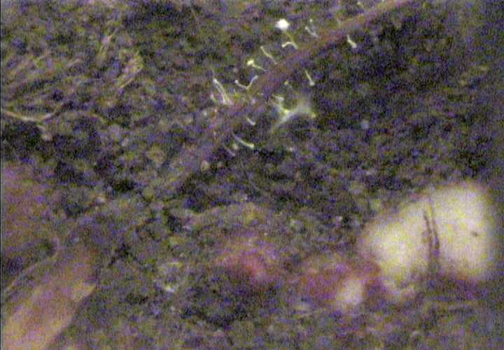
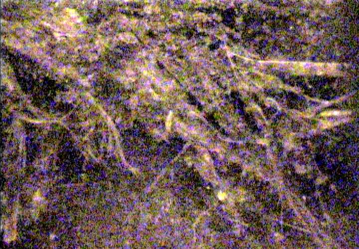
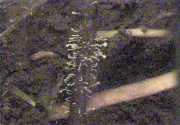
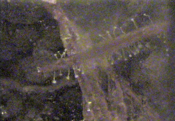
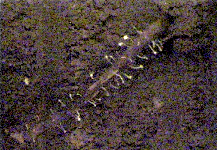
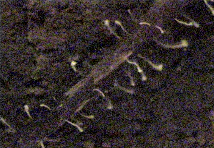
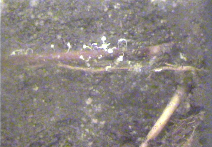
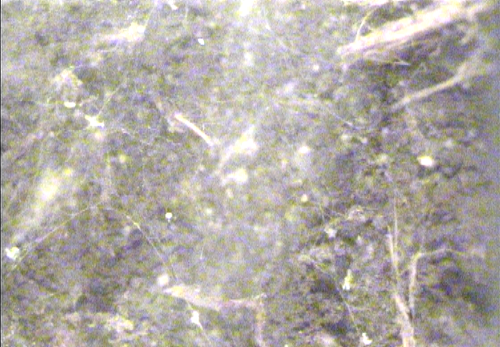
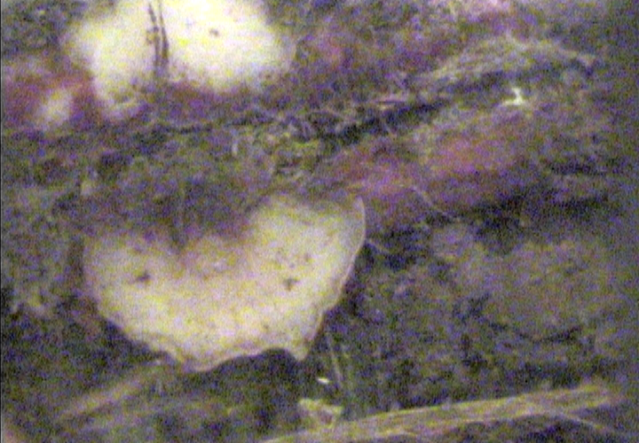
These peculiar structures seem to be fungal fruiting bodies growing
out of the root tissue. You'll see more images below of this strange phenomenon.
|
|
|
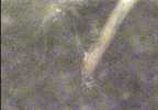
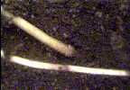
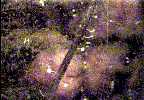
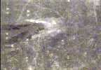
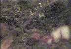
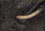
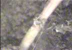
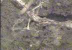
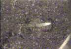
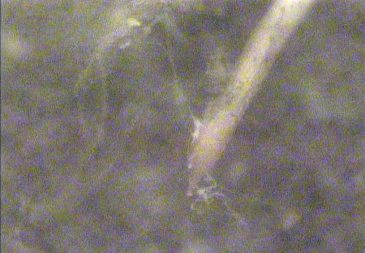
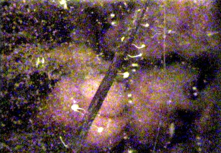
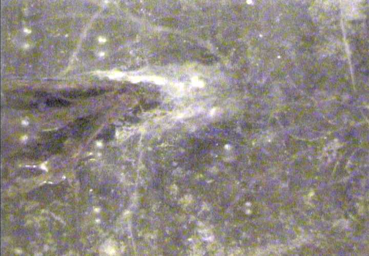
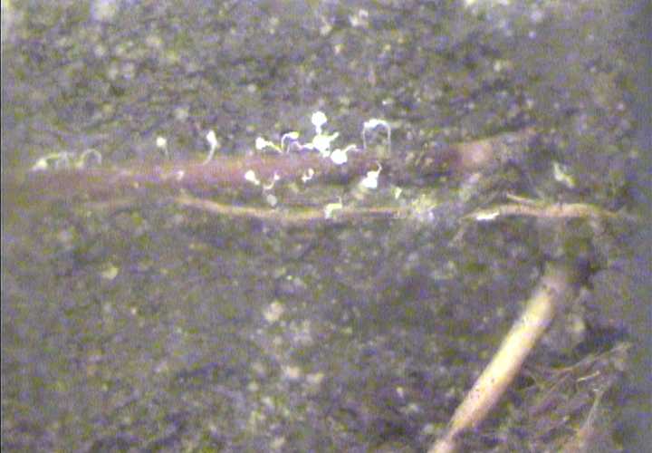
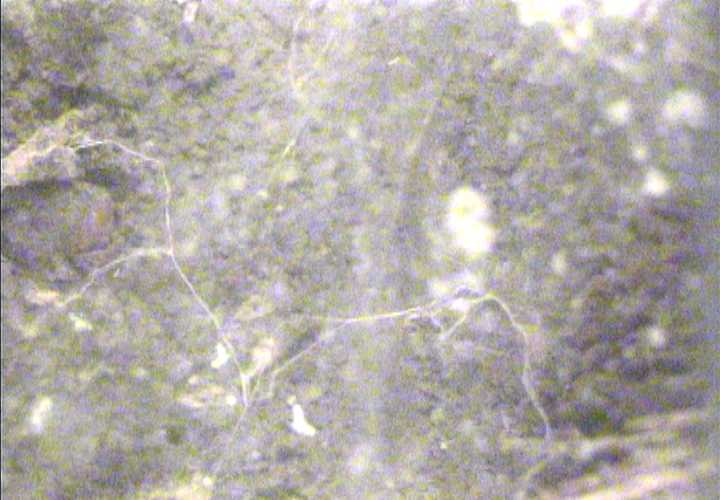
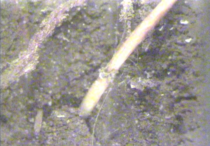
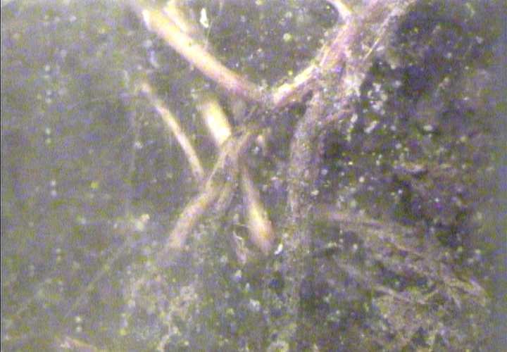
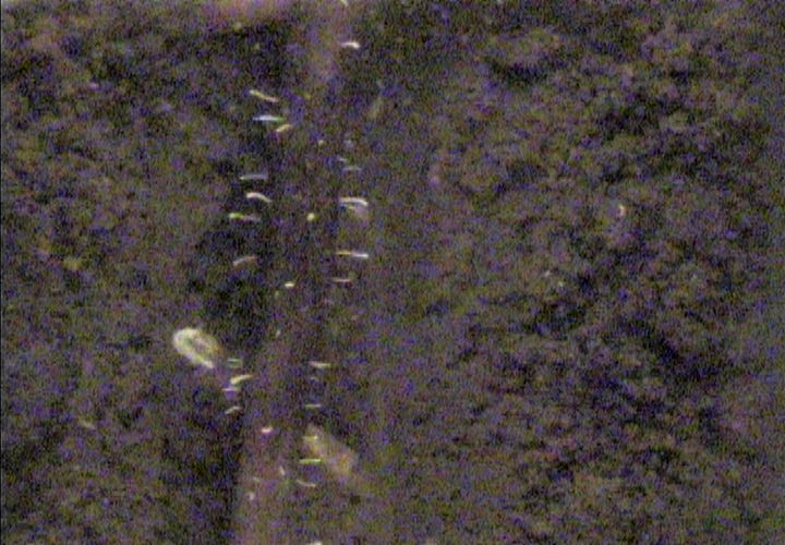
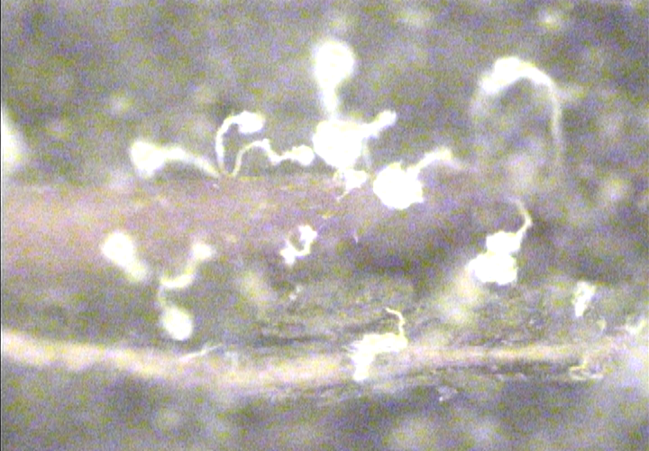
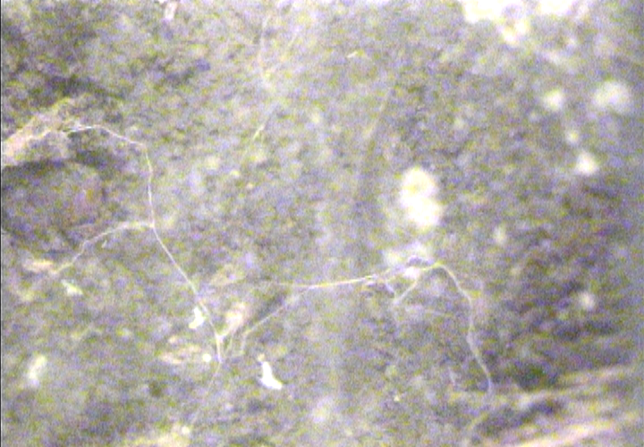
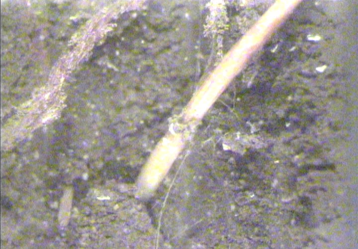
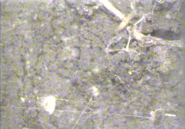
These strands appear to be fungal hyphae infecting a root tip.
|
|
|
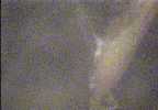
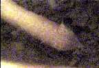
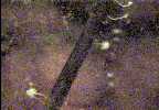
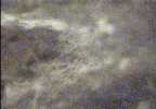
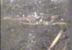
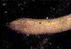
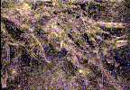
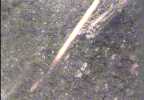
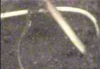
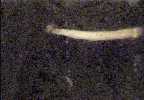
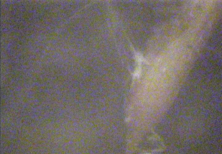
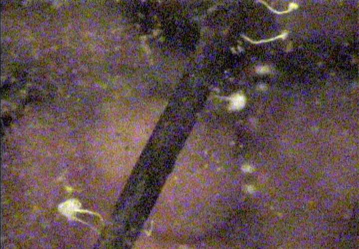
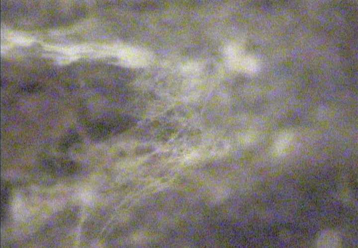
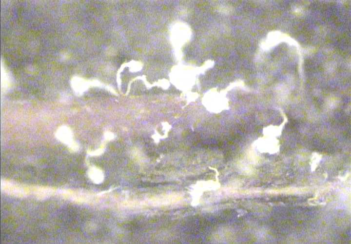
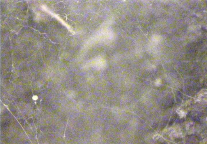
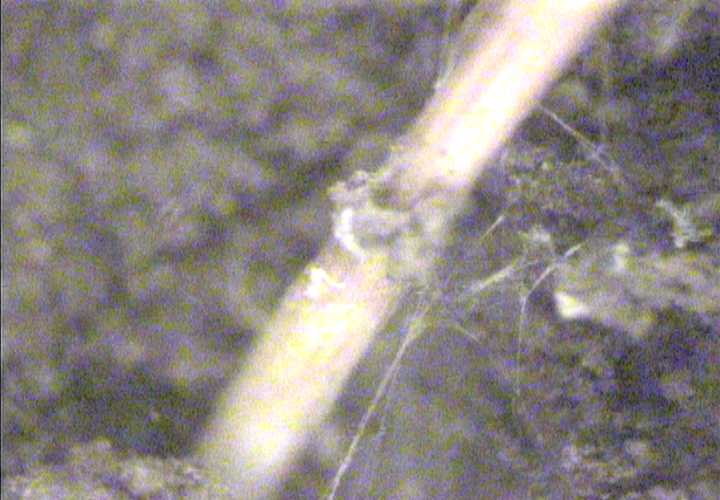
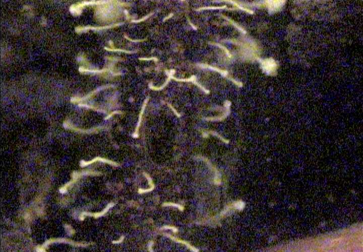
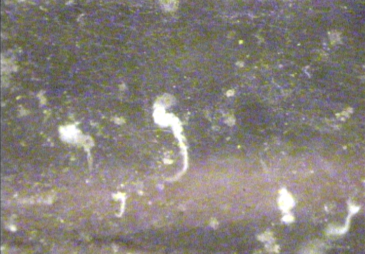
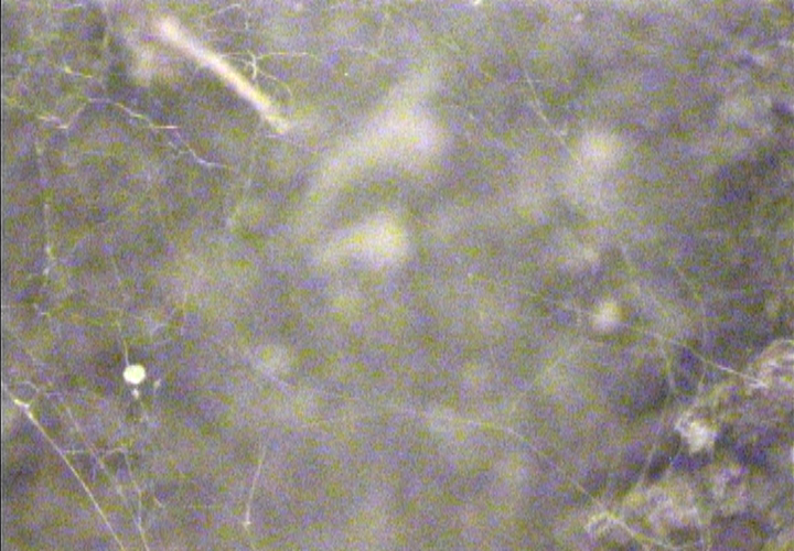
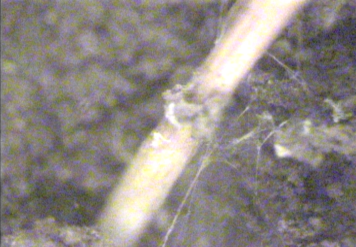
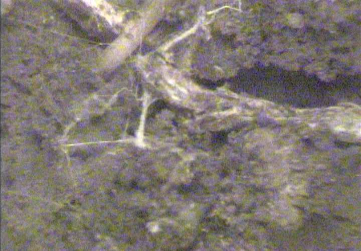
Full zoom on above image...
|
|
|
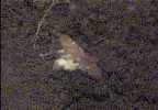
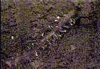
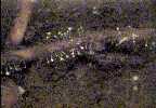
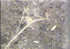
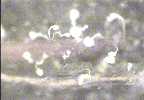
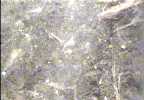
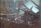
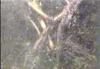
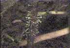
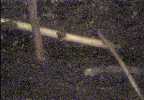
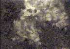
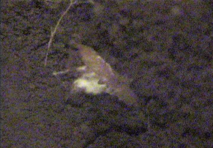
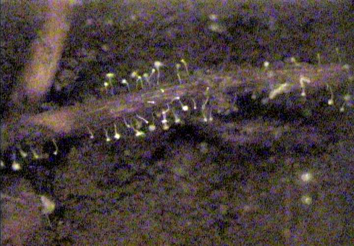
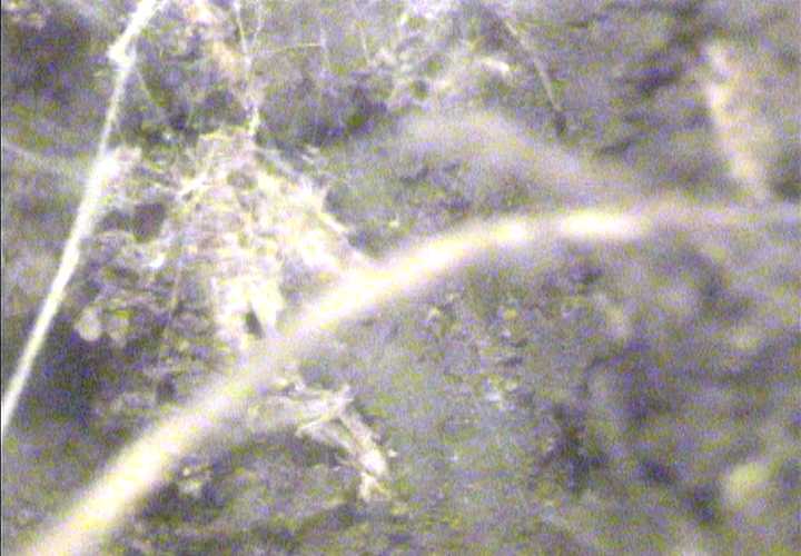
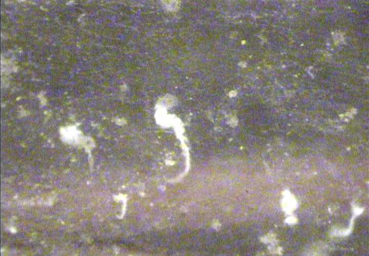
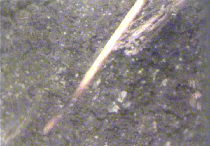
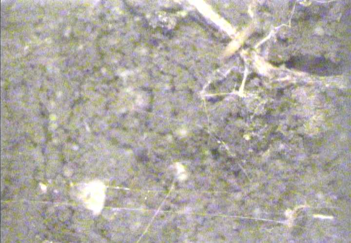
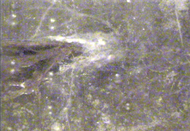
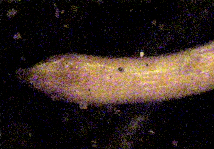
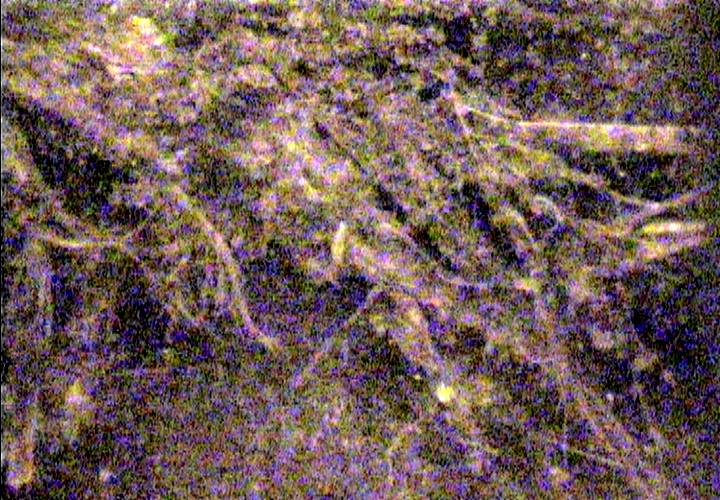
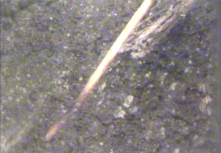
Fungal hot spot; cotton-like...
|

|
|
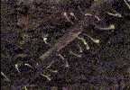
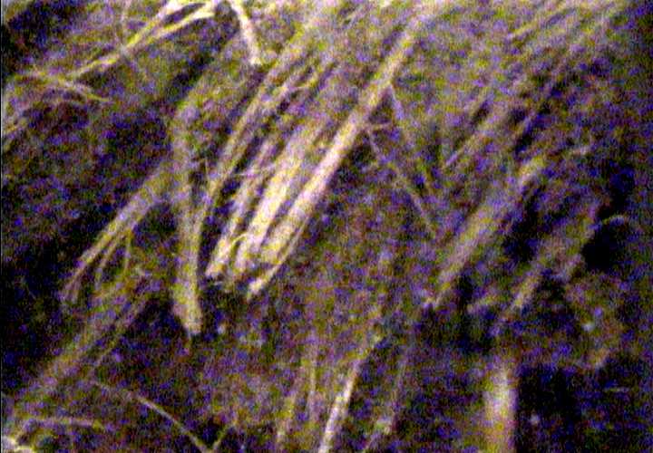
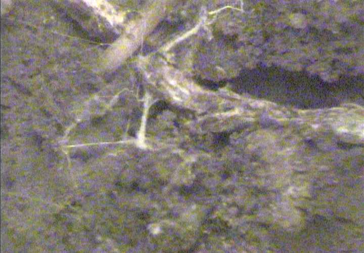
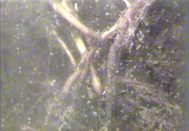
More deep rooting activity.
|

|
|
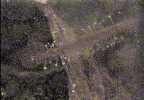
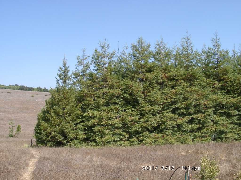
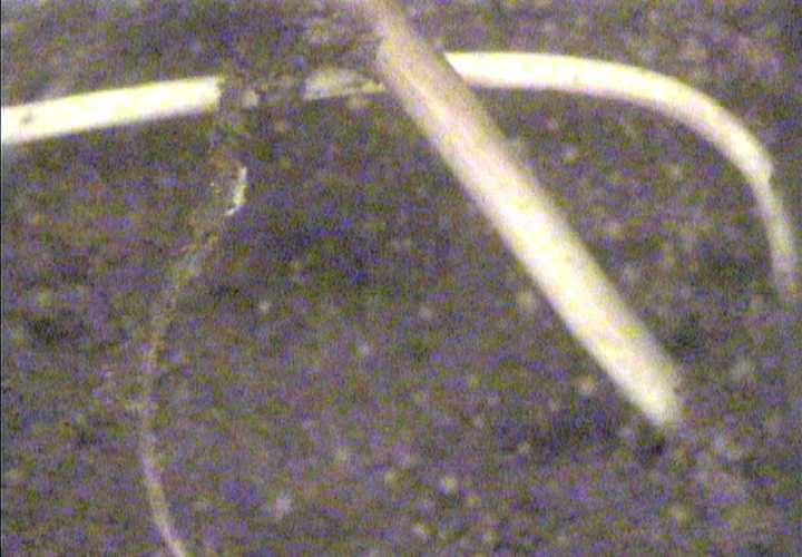
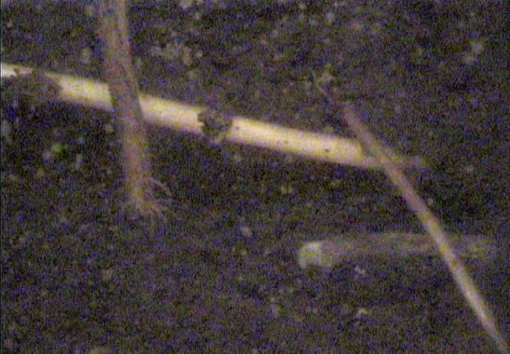
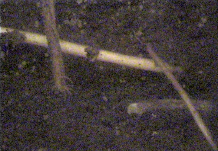
This is a dense root mat. The ragged edge can be attributed to damage incurred
while we augered the hole to bury the tube. Nonetheless, the image shows how extensive
the shallow root systems of S. sempervirens can be.
|
|
|
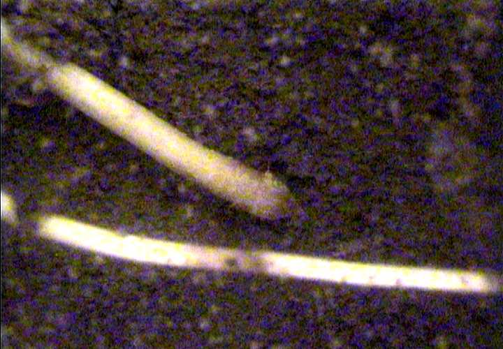
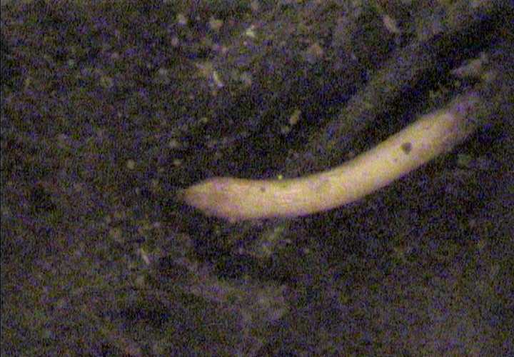
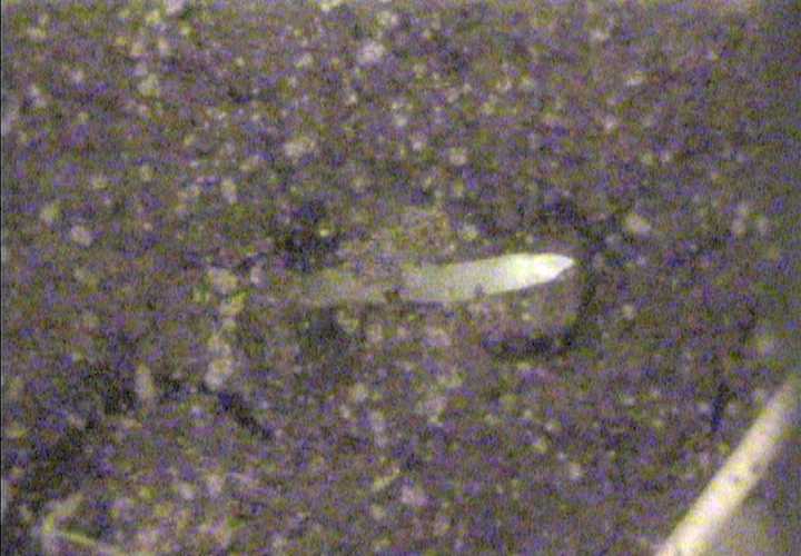
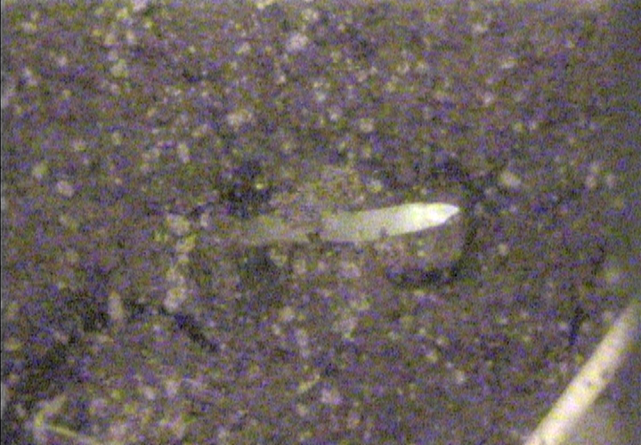
Fleshy root tips...
|
|
|
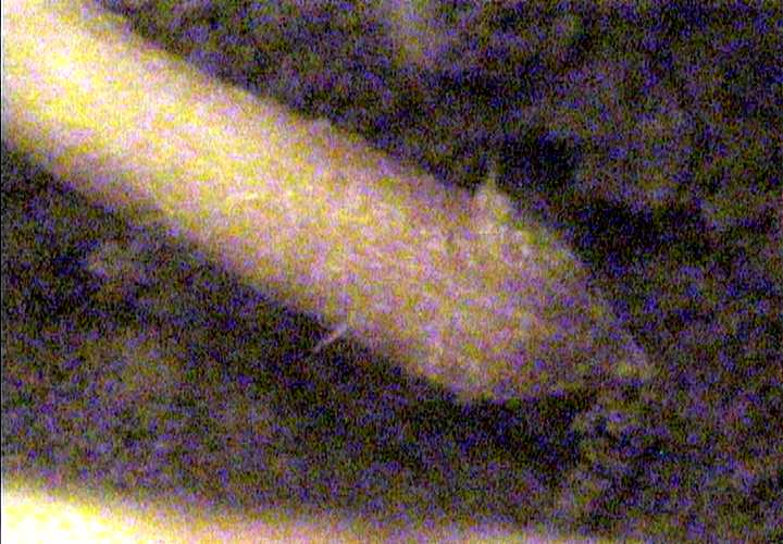
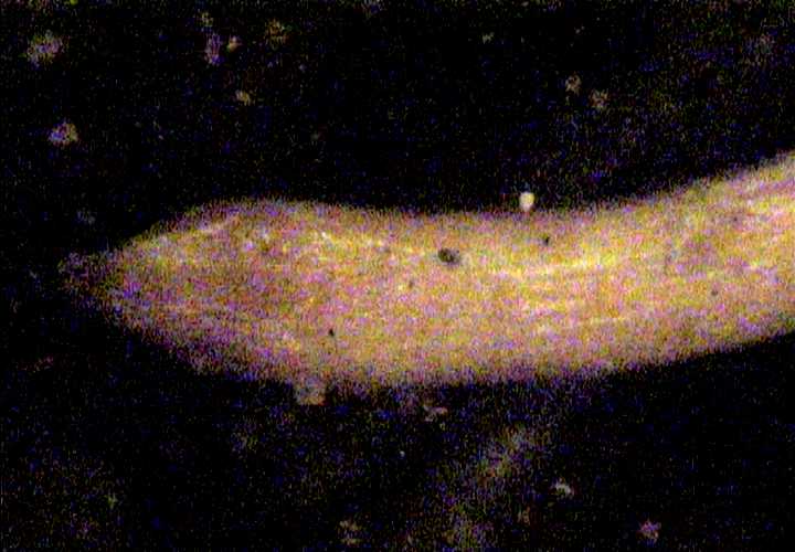
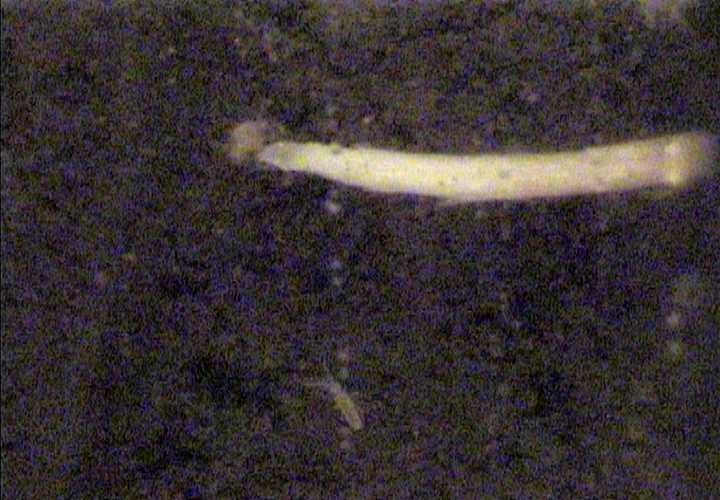
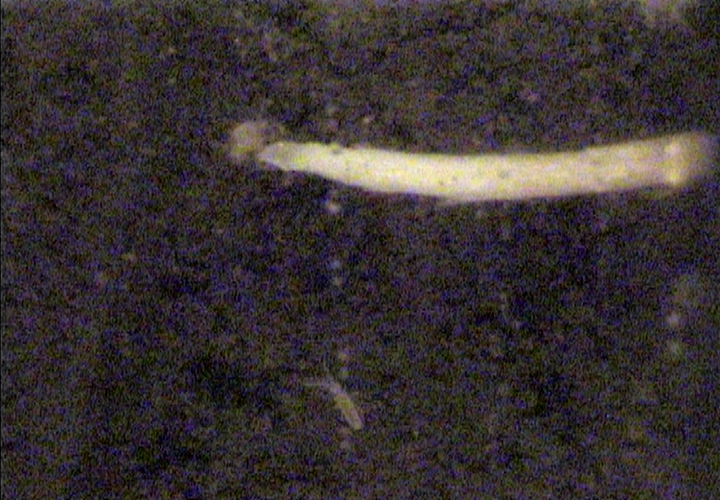
Full zoom on the fat tip from above.
|
|
|
Another fungal-infected root.
|
|
|
Full zoom in on above.
|
|
|
Another fungal-infected root.
|
|
|
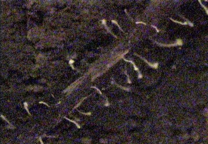
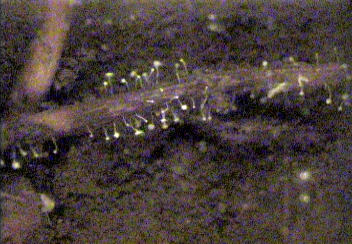
This image nicely shows the shapes of the fruiting bodies.
|
|
|
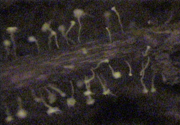
Zoom in on above image...
|
|
|
A 'forest' of fungal branches...
|
|
|
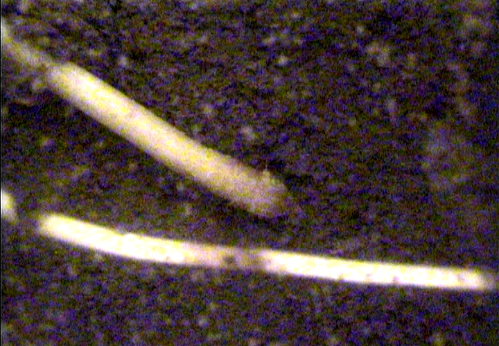
Nice zoom shot of above image; notice the characteristic red color of the
redwood root.
|
|
|
Intense fungal activity. The mycelium appears to have jaws as it consumes
some organic debris in a zone in the soil.
|
|
|
Full zoom on hyphae from the above shot.
|
|
|
The white burst on the screen indicate another fungal hot spot...notice the
insect at the bottom of the screen.
|
|
|
Zoom in on section of mycelia from the above image.
|
|
|
More roots colonized by fungi
|
|
|
Root color contrast and more fungal colonization...
|
|
|
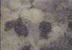
Zoom on image from above gives a good picture of fungal sporocarps/pycnidia.
|
|
|
This zoom illustrates the mycelial network above the spore-producing
structures.
|
|
|
Large fleshy root tip...
|
|
|
Zoom in on above image...
|
|
|
Another fungal hot spot...
|
|
|
More fungal activity; notice the branched hyphae.
|
|
|
Hyphal web...
|
|
|
Dense root mat...
|
|
|
O-horizon; debris and organic matter.
|
|
|
Old and young roots.
|
|
|
Zoom in on young root; notice the web-like fungal hyphae.
|
|
|
2-tone root...
|
|
|
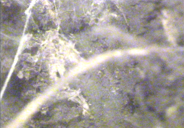
Extensively branched redwood root...
|
|
|
More web-like hyphae associated with roots...
|
|
|
zoom...
|
|
|
Smooth young roots...
|
|
|
Intense fungal activity on older red roots with
unaffected roots in the background.
|
|
|
Zoom on above...
|
|
|
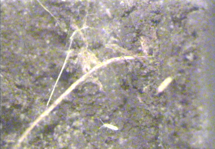
Young growing root tip...
|
|
|
Growing root tip and an arthropod.
|
|
|
Broken roots...possible tube installation effect.
|

|
|
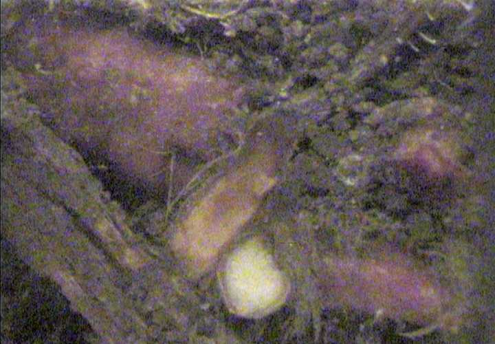
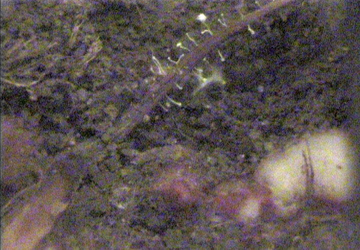
Red bulbous structure like that from the other redwood grove...
|
|
|
Shot #2 of the red bulbous structure...
|
|
|
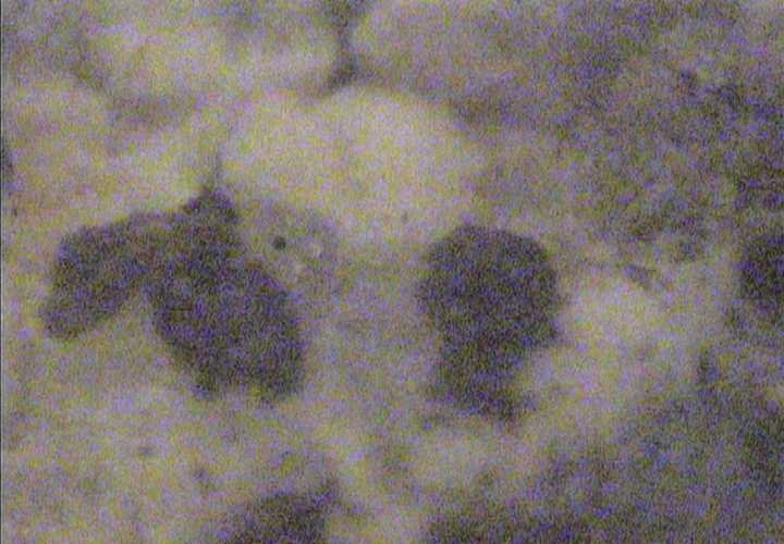
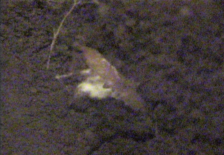
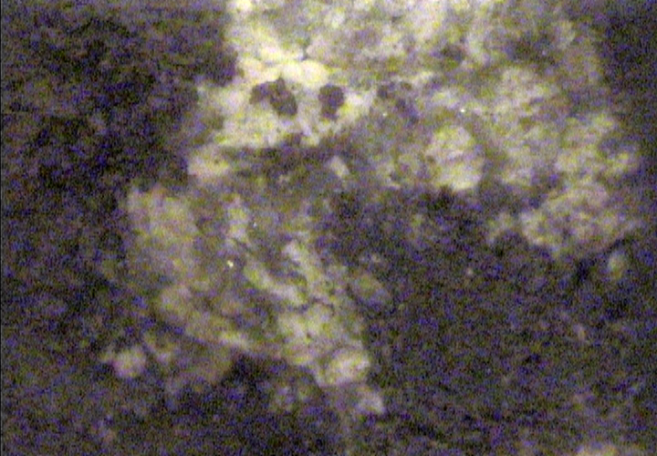
This white mass seems somewhat different from the other white areas of mycelium-rich soil
that we witnessed occasionally. Perhaps this is a different type of fungus?
|
|
|
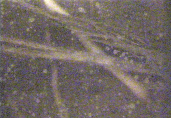
A zoom of the mysterious white substance.
|


